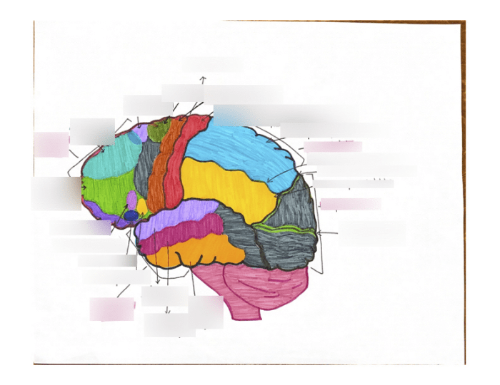
Lesson 6.3 Labeling the Brain: Embark on a journey into the intricate world of the human brain, where we unravel the mysteries of its anatomical structures and delve into the fascinating techniques used to label and study this complex organ.
Through a comprehensive exploration of the brain’s major anatomical structures, we will gain insights into their functions and the crucial role they play in the brain’s overall functioning. We will also discover the significance of labeling the brain for understanding its anatomy and function, examining the various labeling systems employed by researchers and clinicians.
Anatomical Structures of the Brain
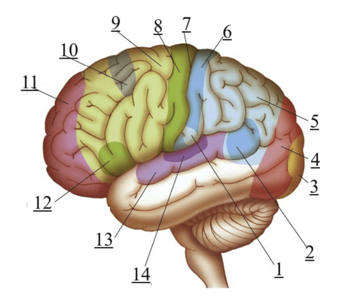
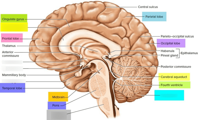
The brain is the control center of the nervous system, responsible for processing information, controlling movement, and regulating bodily functions. It consists of several major anatomical structures, each with its unique functions and contributions to the brain’s overall functioning.
The following table provides an overview of the main anatomical structures of the brain, along with their locations and primary functions:
| Structure | Location | Function |
|---|---|---|
| Cerebrum | Largest part of the brain, located at the front | Responsible for higher-level cognitive functions, including thinking, learning, and memory |
| Cerebellum | Located at the back of the brain, below the cerebrum | Coordinates movement, balance, and posture |
| Brainstem | Connects the cerebrum and cerebellum to the spinal cord | Controls basic life functions, such as breathing, heart rate, and digestion |
| Thalamus | Located at the base of the brain, between the cerebrum and brainstem | Relays sensory information to the cerebrum |
| Hypothalamus | Located below the thalamus | Regulates body temperature, hunger, thirst, and sleep-wake cycles |
| Pituitary Gland | Attached to the base of the hypothalamus | Produces hormones that regulate growth, metabolism, and reproduction |
Labeling the Brain
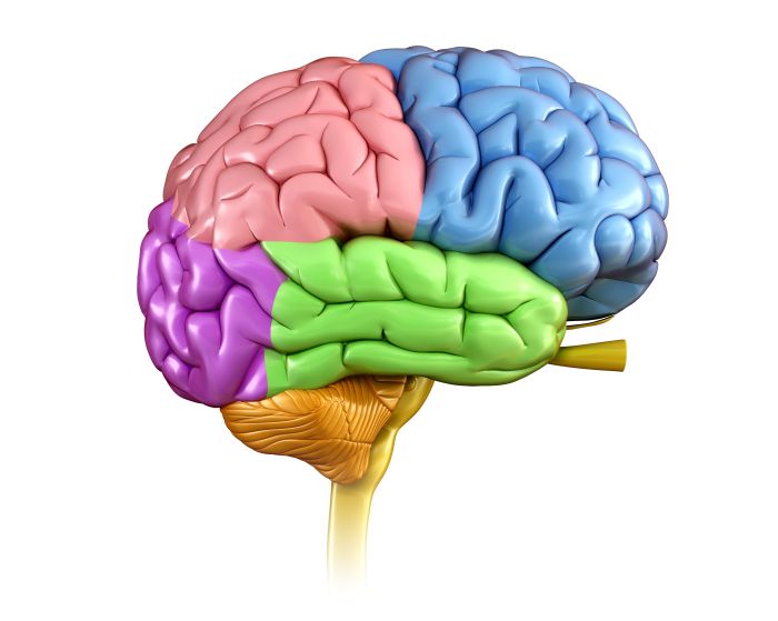
Labeling the brain is crucial for understanding its intricate anatomy and function. By assigning specific names to different brain regions, we can precisely identify and study their roles in various cognitive, emotional, and motor processes.
Labeling Systems for the Brain, Lesson 6.3 labeling the brain
There are several labeling systems used for the brain, each offering unique insights into its organization and function.
- Brodmann areas: This system divides the cerebral cortex into 52 distinct areas based on their cytoarchitecture, the cellular makeup of the tissue.
- Cytoarchitectonic maps: These maps provide detailed descriptions of the different cell types and layers within specific brain regions.
- Functional MRI scans: These scans measure brain activity patterns in response to specific tasks or stimuli, allowing researchers to identify and label regions involved in various cognitive functions.
Benefits of Labeling
Labeling the brain enables researchers and clinicians to:
- Identify and study specific brain regions involved in particular functions, such as memory, attention, or language.
- Compare brain structures across individuals or species to understand variations in anatomy and function.
li>Diagnose and treat neurological disorders by identifying abnormalities in brain structure or activity.
Methods for Labeling the Brain: Lesson 6.3 Labeling The Brain
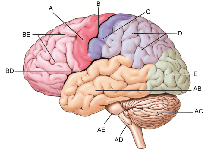
The brain’s intricate structure and vast network of neurons and connections demand precise labeling techniques to study its organization and function. Researchers employ various methods to visualize specific brain regions, cell types, or molecular markers, enabling detailed investigations into the brain’s architecture and activity.
Histological Staining
Histological staining is a traditional labeling technique that involves treating brain tissue with dyes or chemicals to reveal specific cellular components. Different staining methods highlight different aspects of brain anatomy, such as Nissl staining for cell bodies, Golgi staining for neuronal processes, and myelin staining for white matter tracts.
Continuing our study of lesson 6.3, labeling the brain, we’ll explore how these concepts apply to the fascinating topic of há» c bằng lái xe ở mỹ . By understanding the brain’s intricate structure, we gain insights into the complex processes that govern our ability to navigate the world around us.
Returning to lesson 6.3, labeling the brain, we’ll delve deeper into the functions of specific brain regions and their role in our cognitive abilities.
Advantages:
- Widely accessible and relatively inexpensive.
- Provides detailed anatomical information.
Disadvantages:
- Can be time-consuming and labor-intensive.
- Limited specificity and resolution.
Immunohistochemistry
Immunohistochemistry utilizes antibodies to label specific proteins or antigens within the brain tissue. Antibodies are highly specific molecules that bind to target proteins, allowing researchers to visualize their distribution and expression patterns.
Advantages:
- High specificity and sensitivity.
- Allows for multiplexing, enabling simultaneous visualization of multiple targets.
Disadvantages:
- Requires specific antibodies for each target protein.
- Can be technically challenging and expensive.
Molecular Imaging
Molecular imaging techniques, such as magnetic resonance imaging (MRI) and positron emission tomography (PET), use non-invasive methods to visualize molecular processes in the brain. MRI detects changes in water content, while PET measures the distribution of radiolabeled tracers that bind to specific targets.
Advantages:
- Non-invasive and can be performed in vivo.
- Provides functional and metabolic information.
Disadvantages:
- Lower resolution compared to histological methods.
- Can be expensive and requires specialized equipment.
Applications of Brain Labeling
Brain labeling is a crucial technique that finds applications in diverse fields related to the study and treatment of the brain. It has revolutionized our understanding of brain development, disease diagnosis, and surgical planning.In neuroscience, brain labeling helps researchers visualize and map the intricate neural circuitry within the brain.
By labeling specific brain regions or cell types, scientists can study their connectivity, function, and role in behavior. This knowledge is essential for comprehending the complex mechanisms underlying cognitive processes, memory formation, and neurological disorders.In neurology, brain labeling plays a pivotal role in diagnosing and monitoring brain diseases.
For instance, functional magnetic resonance imaging (fMRI) is a non-invasive brain labeling technique that measures brain activity by detecting changes in blood flow. fMRI helps neurologists identify brain regions involved in various cognitive tasks and detect abnormalities associated with conditions such as Alzheimer’s disease and Parkinson’s disease.In
neurosurgery, brain labeling guides surgeons during complex procedures. By using intraoperative MRI or CT scans, surgeons can visualize the labeled brain structures and plan their incisions and resections with greater precision. This reduces the risk of damaging critical brain areas and improves surgical outcomes for patients with brain tumors, vascular malformations, and other neurological conditions.
Top FAQs
What is the purpose of labeling the brain?
Labeling the brain helps researchers and clinicians identify and study specific brain regions, enabling a deeper understanding of the brain’s anatomy and function.
What are the different methods used to label the brain?
The different methods used to label the brain include histological staining, immunohistochemistry, and molecular imaging.
What are the applications of brain labeling?
Brain labeling finds applications in neuroscience, neurology, and neurosurgery, aiding in understanding brain development, disease diagnosis, and surgical planning.
